

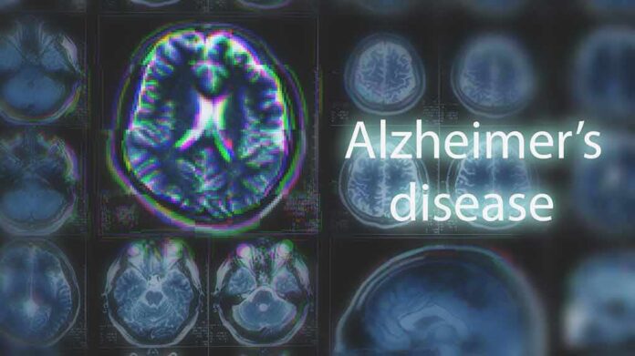
Johns Hopkins researchers say two brain regions may shrink up to a decade before memory loss, raising hopes for earlier Alzheimer’s detection backed by large imaging datasets.
Story Snapshot
- Johns Hopkins teams analyzed hundreds of brain scans from two long-term studies.
- Entorhinal cortex and amygdala showed earlier tissue loss than the hippocampus.
- Changes may appear as much as ten years before memory problems start.
- Finding refines a known imaging path while clinical screening rules remain unchanged.
Johns Hopkins Analysis Flags Earlier Brain Changes
Johns Hopkins researchers examined magnetic resonance imaging and positron emission tomography scans from two independent projects: the Alzheimer’s Disease Neuroimaging Initiative and the Biomarkers of Cognitive Decline Among Normal Individuals. The team reported that the entorhinal cortex and amygdala showed greater relative tissue loss than the hippocampus during the early stages. The group said these changes may be detectable up to ten years before memory symptoms appear, suggesting a tighter early window for risk detection.
The entorhinal cortex helps route memory signals, and the amygdala helps process emotion. Both lie near the hippocampus, which has long been tied to memory loss in Alzheimer’s disease. Earlier studies focused on hippocampal shrinkage as a key magnetic resonance imaging marker. This new work shifts attention to two nearby structures that may change even sooner, refining the map doctors use to study early brain changes in aging adults.
Why This Matters For Families And Doctors
Families fear the day memory fades. Doctors need enough lead time to act. Detecting change sooner could guide who gets closer monitoring, counseling, or future treatments once they are ready. The finding fits a larger push to spot disease years earlier. Clinical guides already view hippocampal atrophy as a major sign on scans, but it is not unique to Alzheimer’s disease. This report adds anatomical focus that could improve how doctors judge risk over time.
Conservatives want tools that work in the real world, not hype that drives costs. Magnetic resonance imaging can be expensive and time-consuming. It needs trained staff and careful setup. Clear, targeted measures can help keep tests efficient and cut waste. If earlier signals in the entorhinal cortex and amygdala prove reliable, doctors could track fewer, sharper markers rather than broad, costly scan batteries for everyone.
How The Finding Fits With Existing Science
Medical literature calls hippocampal shrinkage the most studied magnetic resonance imaging biomarker in Alzheimer’s disease. Average hippocampal volume can drop in mild cognitive impairment and further in dementia. Yet experts also warn that hippocampal loss is not specific to Alzheimer’s disease alone. The Johns Hopkins signal does not replace that record. It builds on it by pointing to neighboring regions that may shift first, offering a better timeline for brain change before symptoms.
Other Alzheimer’s biomarkers include spinal fluid and advanced scans that measure amyloid and tau proteins. Those tools can sharpen diagnosis but add cost and complexity. Imaging remains a required part of dementia workups, according to practical guides, because it also rules out strokes, tumors, or fluid buildup. A more precise structural signal could focus care earlier, while keeping the patient pathway simple and rooted in standard imaging that clinics already use.
What This Means For Policy, Access, And Personal Action
America needs honest health answers without bloated programs. Early, targeted imaging can help people plan care, protect savings, and support loved ones. This research used two respected longitudinal cohorts, which boosts confidence in the signal’s consistency. The report did not publish screening cutoffs or show that this pattern alone predicts future decline for each person, so doctors will still use a full clinical evaluation alongside imaging to guide next steps.
Families can act now. Ask doctors about memory concerns early. Keep records of changes. Discuss whether targeted brain imaging is appropriate. Stay active, eat well, and manage blood pressure and blood sugar, which affect brain health. For policymakers, the priority is clear: support research that makes diagnosis earlier and cheaper, not mandates that add red tape. Strong families and informed patients can drive demand for tools that deliver real value and protect dignity in aging.
Sources:
sciencedaily.com, science.org, medicalxpress.com, nih.gov, nature.com, insideprecisionmedicine.com